Portfolio
A selection of examples of Microscopy, Image, and Data Analysis solutions for Science:
-
Evolution of endocytosis
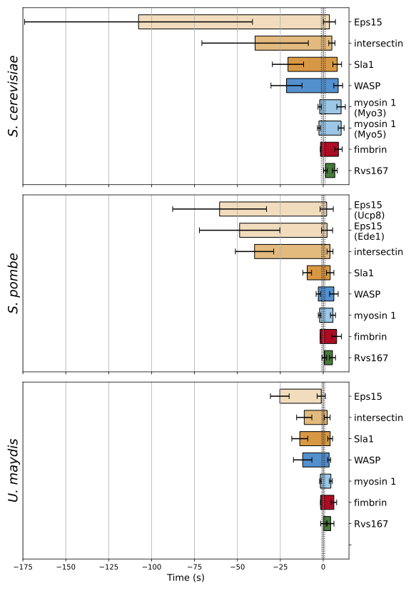
TIRF microscopy enables the precise measurement of the lifetime of endocytic orthologs individually tagged with EGFP in three fungal species: Saccharomyces cerevisiae, Schizosaccharomyces pombe, and Ustilago maydis.
-
Stoichiometry of the ERMES complex
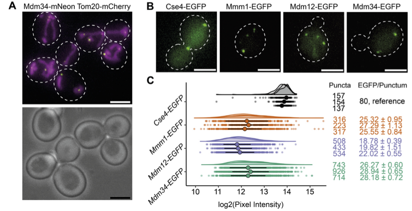
The endoplasmic reticulum–mitochondria encounter structure (ERMES) tethers the endoplasmatic reticulum and mitochondria. It is a four-subunit complex whose stoichiometry and architecture were unknown.
-
Architecture of the exocyst complex
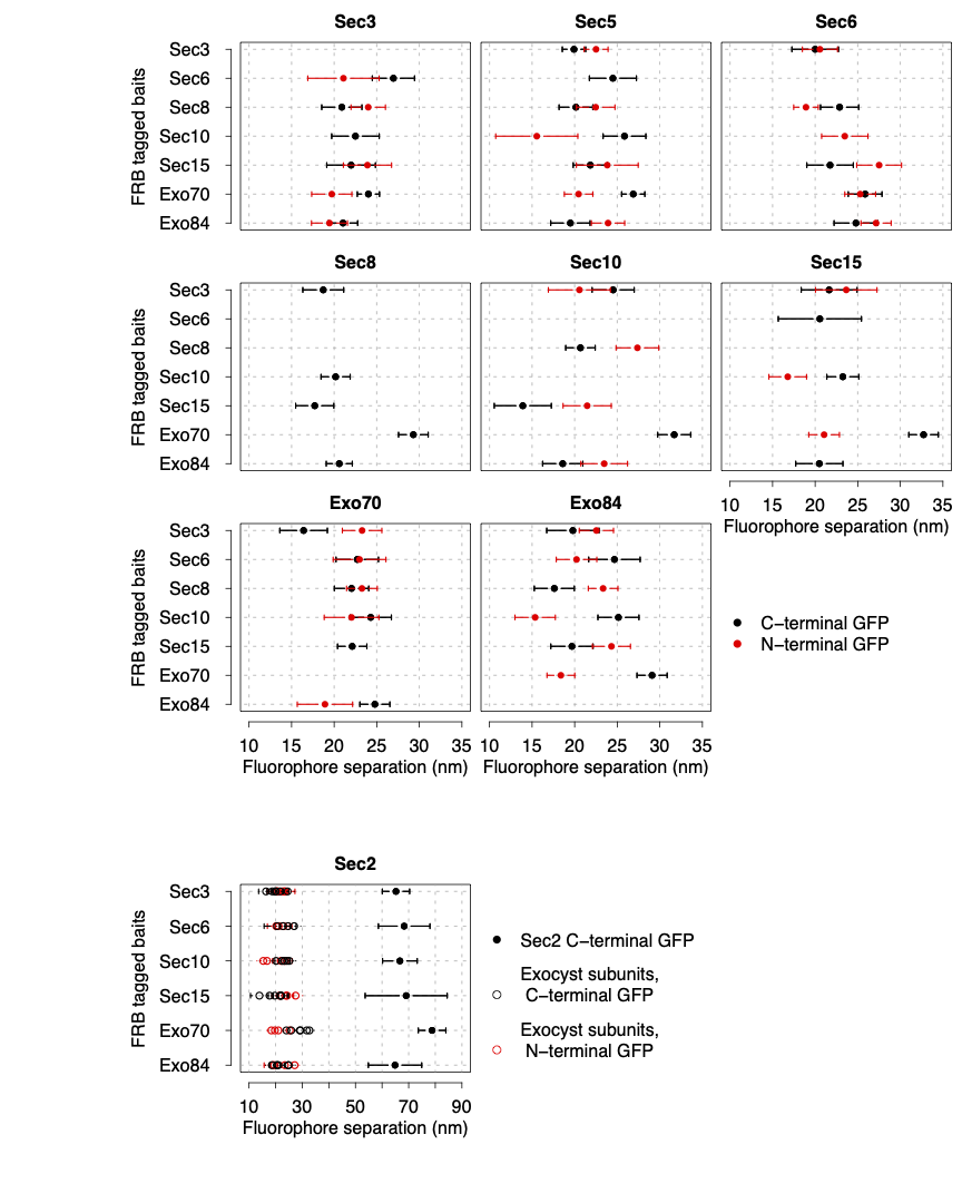
The exocyst is a hetero-octameric complex spanning a few tens of nm. Resolving its protein organisation in living cells would be behind the optical resolution of fluorescence microscopes. We designed an innovative quantitative microscopy approach to trilaterate its architecture with unprecedented precision.
-
Tracking of endocytic dynamics
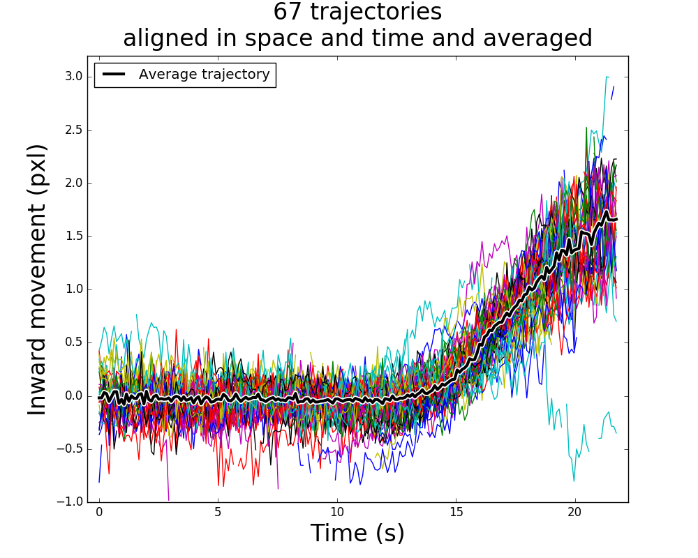
Endocytosis is a very dynamic process where a choreography of hundreds of proteins shapes the cell plasma membrane into an invagination from which a vesicle is released.